


Recent research in the Ocular Surface Laboratory (OSL) has focused on optimising the clinical diagnosis of ocular infestation with Demodex. Typically, Demodex diagnosis involves the (somewhat unpopular) removal of eyelashes and their inspection under a laboratory microscope to count the mites. In situ methods of eyelash manipulation and biomicroscopy, as well as in vivo confocal microscopy have also been proposed, but none of these methods seem to be commonly applied in clinical practice.
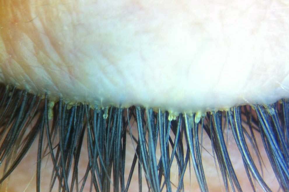
In a recent OSL study, presented as a poster at ARVO 2019 in Vancouver, five established and refined diagnostic techniques were compared and the best of these proposed for routine clinical use. This involved using forceps to remove the cylindrical dandruff around the base of the eyelash to reveal the insertion point of the lash into the lid margin. Next, tension was applied laterally to the eyelash for up to 30 seconds which caused the mite tails to protrude from the follicle orifice, allowing simpler mite identification under the high magnification of a regular slit-lamp biomicroscope.
This newly proposed method was shown to reveal up to four times as many Demodex mites as other reported methods. It appears convenient for both clinicians and patients as it is rapid, painless, requires only basic equipment, and can be repeated at frequent intervals without concerns about lash loss. It is hoped this technique will offer a more reliable, patient-friendly method of gauging the severity of infestation and monitoring the effects of existing and future blepharitis treatments.