


Hosted at a new venue, the Maritime Room, the 9th Glaucoma New Zealand (GNZ) Symposium delivered an excellent programme with loads of great tips and content. The audience of ophthalmologists, optometrists and orthoptists heard updates on treatments, emerging technologies and plenty of discussion on the fundamentals of finding and treating glaucoma.
Keynote speaker Dr Aparna Raniga, Retina Associates Sydney, addressed optometrists, who have a key role in identifying asymptomatic disease. The goal is to maintain quality of life and avoid functional vision loss, which involves looking at the long game in terms of treatment options and duration and side effects, she said. There were reminders about the real challenge of medications and eye drop bottles. In her later talk, Dr Raniga spoke about indications for glaucoma surgery, with an overview of different bleb- and angle-based procedures.
Dr Sam Kain, an honorary senior lecturer at the University of Auckland (UoA), did a superb job as MC and his observations helped create a comfortable space for sharing and learning. Having being introduced as a symposium ingénue, Dr Mia Zhang, a senior glaucoma fellow at UoA, spoke about the limits of eye pressure when looking for glaucoma and considering patients with normal intraocular pressures (IOPs) who are progressing, as well as those with ocular hypertension (OHT) who don’t convert. She encouraged us to be cognisant of a wide range of non-pressure risk factors, from ethnicity to sleep apnoea.
Speakers Drs Hussain Patel, Sonya Bennett, Mia Zhang and Aaron Wong
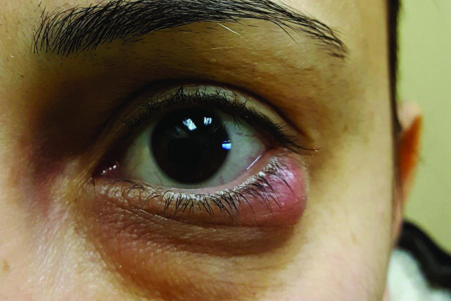
Eye Institute glaucoma specialist Dr Kaliopy Matheos reminded us to always measure colour vision and pupils, while considering conditions that may masquerade as glaucoma. These masqueraders can threaten more than just sight; in an unclear diagnosis there might be a clue in a sentinel event, or the cause might be in the gut with B12 deficiency.
Holding up her gonioscopy lens acquired during her optometry training, Re:Vision’s Dr Divya Perumal had some practical tips for improving our gonioscopy skills and practice. She offered us the acronym ‘SIP’ to follow when looking at angles, where SIP stands for: structures, irido-trabecular contact and a range of conditions comprising plateau iris, pigment dispersion, pseudoexfoliation and other peculiarities. She directed us to the World Glaucoma Association for further gonioscopy resources.

Jas Walia, GNZ's Pippa Martin and Vaishna Singavarothayan
Introduced as a doyen of glaucoma, Eye Doctors’ Dr Mark Donaldson discussed whether normal tension glaucoma (NTG) is a real thing. He assured us it is! Dr Donaldson outlined the dual-pressure theory of glaucoma, intracranial pressure (ICP) versus IOP and the finding that ICP is low in NTG. He reminded us of the features of NTG, such as optic nerve haemorrhages at the inferior rim, lower corneal thickness and people with vasospasm.
Dr Donaldson described undertaking an ‘archaeology’ exercise for these NTG cases; has the pressure been high in the past? Is it previous pigment dispersion syndrome (PDS) that has burnt out? Is there intermittent angle closure? Or a history of steroid use? There was a refresher on proactive tests we can do, such as water drinking, IOP phasing and home IOP monitoring.

Device's Regan Clark with Hamilton Eye's Margaret Petch
One of the gems of this event is a question that throws up clinical pearls. There was an excellent discussion measuring pupil reflexes in eyes with dark coloured irides. Suggestions included subjective brightness testing using a cell phone torch for a brighter light source, holding the light at the bridge of the patient’s nose while doing a cover test, or measuring the pupil size in the slit lamp.
The theme from Nelson’s Dr Antony Suter was how to determine if a patient’s glaucoma is stable. There are various tools available, some of which are in use in everyday practice, such as photographs, ocular coherence tomography (OCT) and visual field index. A high-risk patient with a worse visual field is a situation warranting increasing treatment, he said. When things are progressing despite treatment, the path leads to the next difficult step – surgery. Consideration should be made of patient age and whether the disease is advanced. Dr Suter described late referrals as “depressing work”.

Ava Chen and Robert Ng
OCT was described by Eye Institute’s Dr Graham Reeves as “the new oracle” and something we would be uncomfortable without. He cautioned us, however, to not simply believe what the OCT says. Dr Reeves then took us through the importance of good quality images, how to achieve them and what they mean. Looking at the retinal nerve fibre layer (RNFL) is “where the money is”; however, a good reminder to follow is the ganglion cell analysis, which may show glaucoma earlier, he said.
Patient perspectives and compliance
Amid sessions focused on clinical tests and findings, UoA and Greenlane’s Dr Sonya Bennett’s presentation took a more sombre tone as we considered the burden of disease. It was a good reminder to consider things from the patient’s viewpoint, with associated fear and anxiety around even routine examinations.
Waikato’s Dr Michael Merriman outlined the challenge of managing the growing number of glaucoma suspects. We all have a number of tools at our disposal and have to consider how we manage and follow these patients. There was a call to consider the caseload in our overwhelmed public glaucoma clinics, where confirmed glaucoma patients are also needing follow-up and care.

Sanjay Jairam and Theresa Nowak
Switching from looking for glaucoma, the presentation by Hamilton’s Dr Ben Hoy discussed how to sleuth for compliance. He gave us some cunning tips, such as asking a patient to name their drops, how many times they have missed their drops in the past week and if they have their drops with them. Look at their body language, particularly crossed arms.
If you are finding poor IOPs, be quick to suspect poor compliance, he advised. There are apps to help people both insert the drops and remember to use them, including the EyeDropAlarm and Sydney Eyedrop.

Author Claire McDonald with colleague Emily Kamimura
The case presentations were interactive, with Dr Kain encouraging the audience to be brutal with questions for the panel. I can report that, although presenting a case comes with some nervous moments, the atmosphere was very supportive and there were insights and experiences shared that were great learning. That includes the offside comments by the MC, such as “Google is so helpful in clinic” and, in closing the case session, “It is nice to talk about them (the cases) and see what other people would do – you are on your own a lot.”
The final session of the day has evolved with feedback from previous symposia. GNZ chair Professor Helen Danesh-Meyer presented four cases, and invited the panellists to role-play “What are you going to tell the patient?” to explore that magical balance between a patient understanding the clinical findings without creating undue concern.
Claire McDonald is an optometrist and business co-owner of McDonald Adams Optometrists in Warkworth, north of Auckland, with a post-graduate diploma in science and optometry from the University of Auckland.