


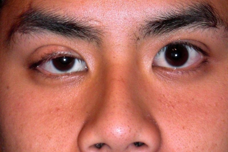
Ptosis is one of those conditions that seems simple until you try to measure it properly. In clinic, between the millimetre ruler, patients shifting their eyes, a lid that twitches at the worst moment and the clinician leaning uncomfortably close to the patient’s eye, the process is far from easy.
Metrics such as margin reflex distance 1 (MRD-1) and palpebral fissure height are essential, but they are also deeply dependent on examiner skill, patient cooperation and lighting. For anxious patients, children or those who dislike close contact, it can be a difficult experience. For clinicians, it is often inconsistent, especially taking measurements over time or between different examiners.
So the question has been hiding in plain sight: why are we doing this manually?
The idea for an alternative solution surfaced at the end of a long oculoplastics clinic, when Dr Cam Loveridge-Easther showed me something he had mocked up using ChatGPT. It was disarmingly simple. The iris is close to a perfect circle. Eyelids obscure predictable arcs of that circle. Therefore, in theory, a computer should be able to measure ptosis from a single photograph by calculating how much of the iris is exposed.
That was the moment the IRIS project began. We decided to test the concept with a small pilot model built in Google Colab, which provides access to GPU (graphics processing unit) computing power for deep learning. My initial instinct was to approach the problem mathematically: treat the pupil as a centre point, fire ‘starburst’ rays outward, detect where they hit the iris or eyelid margins and calculate the amount of obstruction geometrically.
On paper, it worked, but in practice it was chaos. Real photographs have eyelashes, reflections, variable lighting, slight tilts and subtle lid contour changes that break simple geometric rules. It worked adequately in controlled images but fell apart in anything resembling a clinical photograph. It was clear that rule-based systems couldn’t scale. The model needed to learn to recognise eyes the way clinicians do, through exposure to thousands of examples.
To shift to true deep learning, the first step was to use OpenEDS, an anonymous research dataset containing 27,431 labelled infrared eye images. This gave the model anatomical basics, such as where the iris tends to sit and how lids and sclera interact. But infrared images look nothing like the colour photographs we take clinically, so the results didn’t generalise well.
To get closer, I hand-labelled 452 images from UBIRIS, a Brazilian dataset of colour eye photographs taken at a wide range of angles and distances. UBIRIS added the realism of pigmentation, shadows, reflections and the kinds of optical artefacts real eyelids produce. However, we still weren’t sure if these aligned with the conditions of our own oculoplastics service.
The final stage was the most labour-intensive: hand-labelling 250 clinical photographs and using them to test the model. Processed entirely offline to maintain patient confidentiality, these images gave the model the specific teaching it needed to function in real practice.
We measured the model’s performance using intersection-over-union, a standard metric comparing the overlap between the model’s predicted iris and eyelid boundaries and expert annotations. A perfect score is 100%. Our model achieved 94.83%. For context, a recent Scientific Reports study working on eyes without ptosis reported 96.48% accuracy. Considering the variability inherent in ptotic eyelids, our results were extremely encouraging. The reception we got from the RANZCO 2025 Congress reinforced that, with the abstract also selected as an Australian Vision Research notable abstract and the project receiving the Australian & New Zealand Society of Ophthalmic Plastic Surgeons’ for best free paper.
Next steps
There is still work to do. The current version of IRIS requires a user click to identify the approximate pupil centre. We aim to remove that step, creating a fully automatic process from photograph to ptosis estimate. Increasing training volume with more diverse eyelid shapes, ethnicities and photographic conditions should also improve the model further.
We are also planning a clinical correlation study, comparing the AI’s measurements with patient-reported satisfaction before and after ptosis surgery. Ultimately, we want to know whether numerical iris-exposure metrics reflect functional and aesthetic outcomes that matter to patients. Beyond that, the potential use-cases are wide. It could assist in triaging oculoplastic referrals; it could streamline pre-approval processes for insurers, providing reproducible evidence of impairment. It might also support surgical audits, postoperative comparisons and long-term monitoring of progressive ptosis.
The larger point is that AI is not here to replace clinicians, but to extend what we can measure, standardise and interpret. Ptosis assessment is an ideal use-case for AI – it is visually driven, reliant on consistent measurement and currently burdened by manual variability. If an algorithm can deliver fast, objective and reproducible results from a simple photograph, then patients benefit, clinicians save time and the overall quality of care improves.
IRIS is still an early tool, but the current results suggest automated ptosis measurement is both feasible and clinically meaningful. With refinement, we hope to make eye-based AI accessible, reliable and integrated into everyday oculoplastic practice.
James Lewis is a fifth-year ophthalmology registrar at Capital & Coast DHB. His work sits at the intersection of ophthalmology, AI and health-system efficiency. He is a published author with awards, including the 2023 Palmerston North Post-Graduate Medical Society RMO Prize for his NZMJ paper on the health economics of faricimab.