


Researchers in Australia found eyecare professionals grading retinal photographs with a structured visual search strategy diagnosed diabetic retinopathy (DR) with higher accuracy than those scanning for regions with prominent features.
Researchers at Deakin University recruited 15 optometrists and eight fellowship-trained ophthalmologists, who were given 40 retinal images to grade for DR while their eye movements were recorded using a Gazepoint GP3 device. The retinal images were taken from the publicly available Chinese DDR dataset, which are classified according to DR severity: none, mild, moderate, severe or proliferative DR.
Analysis showed differences in gaze behaviour between correct and incorrect respondents (‘incorrect’ being any grade not matching the image’s recorded grading). Notably, while optometrists had higher fixation counts, total visits per area of interest (AOI) and total time spent at each AOI, compared to ophthalmologists, no difference was found between the two professions’ grading accuracy. However, optometrists showed comparably lower accuracy in detecting proliferative DR, said researchers.
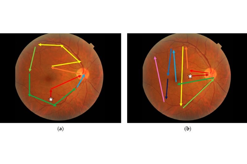
The pattern associated with correct grading (see picture) shows little redundancy and perhaps indicates a conscious strategy of starting at the macula before looking around the superior arcade, then around to the inferior arcade, finishing at the optic disc, said researchers, writing in the Journal of Clinical Medicine. “The pattern associated with incorrect response starts in the nasal region and moves temporally. This may suggest that a structured search pattern based on the anatomical features of the retina is superior to a saliency-based search when grading diabetic retinopathy from fundus photographs.”
The study suggests referable DR is detected at high rates but with disagreement between clinicians when determining a precise severity grade, concluded researchers. “While it may be possible to instruct clinicians on the search patterns that they should use, work by Kok et al. (2017) showed this must be paired with knowledge of the underlying condition to improve clinical performance. Hence, an intervention looking to change visual search behaviours should also involve a targeted educational component.”