


Infestation of the eyelash follicles and meibomian glands with Demodex mites is emerging as a significant cause of blepharitis. This article reviews key aspects of Demodex biology and pathogenesis, as well as the epidemiology, clinical manifestation, diagnosis and treatment of infestation.
Demodex biology and epidemiology
Two species of Demodex infest the human eye: Demodex folliculorum inhabit the eyelash follicles, while Demodex brevis reside in the meibomian glands. These species are therefore capable of causing both anterior and posterior blepharitis, respectively (1). The larger of the two species, D. folliculorum grows up to 0.4mm in length, while D. brevis are typically up to 0.2 mm long. As D. folliculorum are found close to the opening of the eyelash follicle, frequently with the posterior end of the abdomen protruding and visible, while D. brevis are located deep in the meibomian glands, the transmission of D. folliculorum is higher and has become recognised as the most common parasitic infestation of the eyelid (2, 3). The reported life span of Demodex is variable, with estimates of 28 and 100 days, however, mite survival is limited outside the human body (1, 4, 5). Transmission occurs via direct contact and transfer of the adult mites, larvae or eggs.
The prevalence of Demodex infestation rises with age, with reported infestation rates of 13% in individuals 3-15 years of age, 69% in those 31-50 years of age and over 95% in individuals aged 71 years or older (6). Demodex load in affected individuals also increases with age. Ocular demodicosis is associated with rosacea - the presence of Demodex in the hair follicles and sebaceous glands of the face results in the development of rosacea and it is postulated that these mites spread to the eyelids, leading to periocular infestation (7).
Clinical manifestation
The presence of Demodex is strongly associated with the development and exacerbation of blepharitis, with mites detected in up to 68% of patients with chronic blepharitis (8). Cylindrical crusting around the eyelashes is considered pathognomonic for demodectic blepharitis (2). This crusting is believed to comprise debris from damaged epithelial cells, by-products of meibum digestion and mite excreta. Demodex has been detected in 60% of those with meibomian gland dysfunction (MGD) (9), the most common form of posterior blepharitis that affects the delivery of lipid to the surface of the tear film.
Characterised by inflammation of the eyelids, blepharitis is one of the most prevalent ocular conditions worldwide, affecting around 47% of patients seen by eye health professionals. In more severe cases, it can result in the development of more serious disorders such as eyelid infection, cyst formation and sight threatening corneal sequelae. Irritated dry eyes, intermittent visual disturbance, and ocular surface and eyelid redness, however, are symptomatic of blepharitis at all levels of severity.
Of note, Demodex is also common in asymptomatic individuals – up to 18% of those affected do not exhibit symptoms – and it is uncertain why the presence of Demodex elicits an adverse reaction in some individuals, while others remain unaffected. It may be that Demodex constitutes part of the normal skin flora, and that immunodeficiency may be associated with the development of symptoms, although this remains to be established (10-12). Also unclear is the extent to which mite load correlates with symptom severity. Further investigation into factors affecting disease development is clearly needed.
Pathogenesis
Consumption of the epithelial cells of the eyelash follicle and meibum-producing cells of the meibomian gland, together with blockage of the eyelash follicle and meibomian glands by Demodex may lead to madarosis and trichiasis, as well as insufficient meibum secretion, respectively. The sharp chelicera (appendages such as the mouthparts and claws) of Demodex may also cause epithelial damage, leading to epithelial hyperplasia, hyperkeratosis and crusting. Demodex may also directly and indirectly trigger immune hyperstimulation. Mite excreta as well as chitin, the main component of the Demodex exoskeleton, are antigenic, as are by-products from their digestion of meibum. In addition, Demodex is viewed as a potential vector for bacteria, particularly Bacillus oleronius, which further triggers immune responses.
Diagnosis
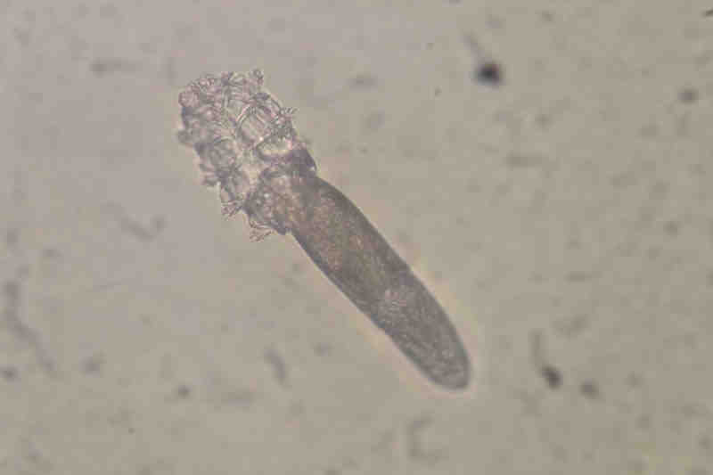
As previously mentioned, the observation of cylindrical crusting encircling the eyelashes under slit lamp biomicroscopy may be a convenient clinical indicator of ocular demodicosis (2). However, the current gold standard diagnostic test remains the examination of epilated eyelashes for the presence of mites, under 250x magnification light microscopy. It has been suggested that targeted epilation of eyelashes with cylindrical crusting and up to 30 seconds rotation of the eyelash within the follicle prior to epilation may increase Demodex yield (2, 13). However, this diagnostic approach is not without challenges; many mites can remain inside the follicle following epilation, mite numbers on each eyelash can vary considerably, and mites may be obscured by crusting and debris. Finally, due to the inaccessibility of Demodex residing deep in the meibomian glands, infestation in cases of posterior demodectic blepharitis remains difficult to diagnose.
The utility of in vivo confocal microscopy (IVCM) and polymerase chain reaction (PCR) in the diagnosis of ocular demodicosis are being explored. IVCM can permit visualisation of Demodex inside the eyelash follicle without epilation; its ability to assess mites within the meibomian glands in cases of posterior demodectic blepharitis should also be investigated (14). In the laboratory, PCR can be used to detect minute quantities of genetic material from Demodex. The Ocular Surface Laboratory at the University of Auckland is currently developing PCR-based and other molecular diagnostic tests in the hope of improving the diagnosis of ocular infestation.
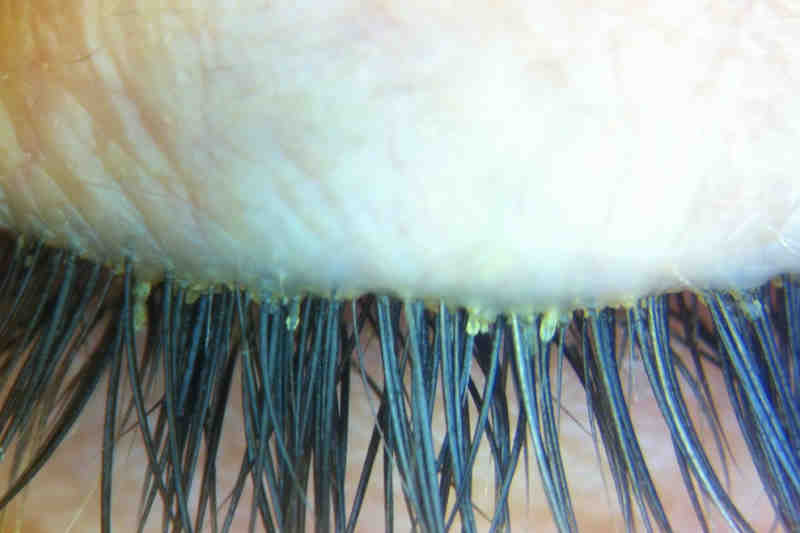
Figure 2. Cylindrical eyelash crusting under slit lamp biomicroscopy is considered pathognomonic of Demodex infestation.
Treatment
Demodex are susceptible neither to antibiotic therapy nor to conventional blepharitis treatments such as eyelid hygiene, artificial tears and warm compresses. Tea tree oil (TTO) is the current standard treatment for ocular demodicosis to target the causative organism. Extracted from the Melaleuca alternifolia tree, TTO has a long history of use for its antiseptic properties. Topical treatment of the eyelid region with TTO decreases Demodex load, ocular surface discomfort, tear film abnormalities and ocular inflammation and improves visual acuity (9). A regime of 50% TTO weekly (mixed with a bland oil such as almond or macademia oil) followed by cleansing with a 10% TTO solution daily for 4 weeks has been described (15), as has a regime of 50% TTO weekly combined with TTO shampoo daily for 6 weeks (16). Ocular surface irritation induced by full strength TTO precludes its use clinically. Lower concentrations of TTO have also shown some benefit (17). Terpinen-4-ol is the key active component of TTO and exhibits an acaricidal effect on Demodex. Specialised eyelid cleansers, containing TTO and terpinen-4-ol to facilitate eyelid hygiene in the management of transmissible blepharitis, are commercially available. However, their efficacy in managing demodectic blepharitis has not yet been confirmed in the literature. Studies are underway at the Ocular Surface Laboratory to assess and compare levels of terpinen-4-ol in commercial eyelid cleansing preparations and their relative effects on Demodex viability in vitro.
As both TTO and acaricides have adverse effects on the ocular surface, the Ocular Surface Laboratory is also currently investigating the efficacy of a topical cyclodextrin-complexed Manuka honey preparation on Demodex viability in vitro and on ocular demodicosis in vivo.
Less commonly, systemic treatments are prescribed to manage Demodex infestation. Oral ivermectin (used in sheep dip), prescribed alone or in combination with metronidazole, has been shown to reduce Demodex load and the clinical signs and symptoms of infestation, with a greater improvement demonstrated by the combination therapy (18).
References
Dr Isabella Cheung is a research fellow in the Department of Ophthalmology at the University of Auckland.