A protocol for reliable dry eye diagnosis
Dry eye disease significantly impacts patient wellbeing worldwide1,2. Tear meniscus height (TMH) is a critical aetiological indicator of aqueous-deficient dry eye, with TFOS DEWS II guidelines defining <0.20mm as the deficiency threshold3,4. Clinical TMH measurement reliability could vary according to the location of image capture along the lower lid margin, or to the timing of assessment relative to a blink, or to the illumination used5. To address these potential inconsistencies, we, together with OSL's Professor Jennifer Craig during her research visit to Aston University, aimed to optimise a standardised protocol using digital video imaging and image analysis6.
Methods
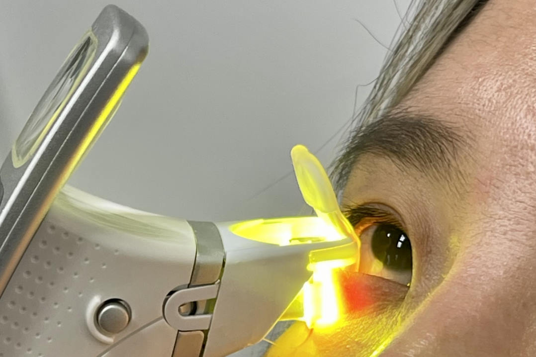
A total of 38 healthy adults (mean age 33 ± 10.6 years; 45% male) were evaluated with the Oculus Keratograph 5M6. After two non-forced blinks, the lower tear meniscus was video recorded for five seconds, under infrared light, then white light. This was repeated twice. Participants also completed the Ocular Surface Disease Index (OSDI), and tear film stability was assessed via three averaged non-invasive keratograph breakup time (NIKBUT) measurements5,6.

Fig 1. TMH measurements using infrared (A) and white light (B) imaging. Yellow markings show measurement points along the temporal, centre and nasal regions of the lower eyelid
For the meniscus height assessments, still frames were extracted at 0.5 second intervals following two successive blinks, for the five seconds of each recording. TMH was measured at seven predefined locations along the lower eyelid margin using the ImageJ software analysis tool, centrally beneath the pupil, then 1, 3 and 6mm nasally and temporally, as shown in (Fig 1). A single trained observer performed all measurements.
Results
TMH measured slightly greater in magnitude under infrared light (0.29 ± 0.08 mm) than with white light (0.27 ± 0.08 mm). This difference is clinically insignificant and aligns with prior reports5. TMH was consistent during the 1.0–2.5 second period after each blink, defining the optimal measurement window6. Taking the measurement directly below the pupil centre and within 1mm nasally or temporally (pupil centre ± 1mm) yielded similarly reliable values, while measurements at peripheral locations showed greater variability6.
No significant relationship emerged between TMH and age, sex, OSDI scores or NIKBUT6. Notably, 34% of healthy participants met the TFOS DEWS II dry eye criteria for aqueous deficiency³. TMH distribution was <0.1mm (0.7%), 0.1–0.2mm (16.9%), 0.21–0.3mm (50.1%), 0.31–0.4mm (24.2%), >0.4mm (8.0%)⁶. A small but statistically significant TMH increase occurred with repeated measurements6.
Conclusion
The protocol we optimised as a proxy for tear volume provides a dependable means of evaluating tear meniscus height6. Imaging centrally on the lid margin, within 1.0–2.5 seconds after the blink and under consistent illumination reduces variability3,5,6. Only a single measurement from a single tear meniscus image is required. The manual protocol establishes a clinical benchmark for subjective or objective analysis of TMH5.
Clinical implications
Implementing this protocol has the potential to deliver:
- Improved diagnostic reliability for aqueous deficiency3,6
- Objective treatment monitoring (eg. following punctal plug insertion)
- Enhanced inter-clinician consistency5,6.
Key points
- Either white or infrared light can be used for imaging or observation, as long as it is consistent
- The measurement should be taken 1.0–2.5s after two consecutive blinks
- The height of the tear meniscus should be assessed within ± 1mm of pupil centre, perpendicular to the eyelid margin and with only a single measurement required5,6
- Staff training can help to identify and exclude artefacts6 .
Automated analysis tools reduce subjectivity and enable large-scale screening2,8. Adopting these protocols helps bridge the research-clinical gap to improve dry eye management1,7.
References
- Dash N, Choudhury D. Dry Eye Disease: An Update on Changing Perspectives on Causes, Diagnosis, and Management. Cureus. 2024;16(5):e59985. Published 2024 May 9. doi:10.7759/cureus.59985
- Stapleton F, Alves M, Bunya VY, et al. TFOS DEWS II Epidemiology Report. Ocul Surf. 2017;15(3):334-365
- Wolffsohn JS, Arita R, Chalmers R, et al. TFOS DEWS II Diagnostic Methodology report. Ocul Surf. 2017;15(3):539-574
- Pena-Verdeal H, Garcia-Queiruga J, Sabucedo-Villamarin B, Garcia-Resua C, Giraldez MJ, Yebra-Pimentel E. A Comprehensive Study on Tear Meniscus Height Inter-Eye Differences in Aqueous Deficient Dry Eye Diagnosis. J Clin Med. 2024;13(3):659. Published 2024 Jan 23.
- Soares I, Ramalho E, Brardo FM, Nunes AF. Tear meniscus height agreement and reproducibility between two corneal topographers and spectral-domain optical coherence tomography. Clin Exp Optom. 2025;108(4):430-436
- Wolffsohn JS, Ayaz M, Bandlitz S, von der Höh F, Ebner A, Craig JP. Optimising the methodology for assessing tear meniscus height using digital imaging. Cont Lens Anterior Eye. 2025;48(4):102419.

Professor James Wolffsohn is head of the School of Optometry at Aston University, UK, and an honorary professor in the University of Auckland’s department of ophthalmology. His main research areas are the development and evaluation of ophthalmic instrumentation, contact lenses, intraocular lenses and the tear film.

Moonisah Ayaz is a research coordinator at the School of Optometry, Aston University, Birmingham, UK.


