


Glaucoma New Zealand’s symposium can always be relied upon to provide an engaging day of education. On a fine morning after an early start (the clocks had sprung forward to daylight savings time), the new venue at Ellerslie Racecourse offered an ideal setting.
This year’s keynote address came remotely from the University of Melbourne and Centre for Eye Research Australia’s Dr Flora Hui. She updated us on research into nicotinamide (vitamin B3) as an adjunct therapy for glaucoma. One theory of how it contributes to ganglion cell health involves metabolic failure’s role in glaucoma. Nicotinamide is a precursor to nicotinamide adenine dinucleotide (NAD), which is vital to mitochondrial capacity to provide energy to hungry ganglion cells (see our January 2022 story). As NAD levels in the retina fall with age, nicotinamide supplements could compensate. However, as Dr Hui pointed out, it’s hard to measure neuroprotection.
Patients are being recruited for a study involving daily doses of 750mg nicotinamide from the Insolar dietary supplement. There is no evidence so far that a supplement could delay glaucoma onset and, disappointingly, my daily spread of Marmite, while containing a mighty dose of B12, does not contain nicotinamide!
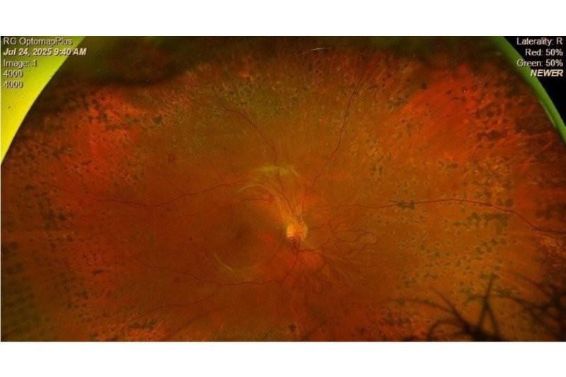
We had some useful reminders from the University of Auckland’s Dr Hussain Patel about the importance of optic disc evaluation to underpin a glaucoma examination. Starting with pragmatic advice to dilate when needed, and to slow down, Dr Patel then ran through essential checks of disc size, the rim, and use of the Disc-Damage Likelihood Scale, a valuable tool. In closing, he suggested we monitor tricky discs in the context of other clinical findings.
A field day
Ophthalmologist Dr Alex Buller from the Eye Surgery Hastings always has a reliably practical approach, this time covering visual field progression. He acknowledged that although no one likes visual field tests, they are the only visual assessment to show glaucoma progression. There were useful reminders to prioritise central visual field tests (keeping in mind that inferior field loss is worse for a patient) and be prepared to follow a suspicious field for some years, if necessary, to get past any noise in the testing. Dr Buller described the logistical nightmare of monitoring visual fields in shared care, mentioning approaches including the Hodapp scale, along with the global glaucoma staging system. The reminder that glaucoma is not always a linear disease stood out for me.
A quote from the director emeritus of the Glaucoma Service at Wills Eye Hospital, Dr George Speath, “You only see what you look for; you only look for what we know,” led into a list of ‘confessions’ from the University of Auckland’s (UoA) Professor Helen Danesh-Meyer. In the spirit of the day, her talk was all about sharing knowledge and experience. Discussing the importance of patient education, she encouraged us to give them a perspective on their glaucoma, reminding us that an increase in patient knowledge can decrease anxiety. Covering many of the tricky aspects of glaucoma care, Prof Danesh-Meyer reminded us that visual field loss does cause symptoms, and once a field is lost, we’re not getting it back.
Prof Danesh-Meyer’s mention of selective laser trabeculoplasty (SLT) as a primary therapy segued nicely into UoA’s Dr Jay Meyer’s presentation. He described the procedure and referenced the laser in glaucoma and ocular hypertension (LiGHT) study, which compared SLT to treatment with drops. After three years, around three-quarters of the SLT group were no longer on drops. Interestingly, none had proceeded to glaucoma surgery. The UK’s National Centre for Health and Care Excellence (NICE) lists SLT as a first-line treatment, as does the American Academy of Ophthalmology (AAO) and European Glaucoma Society (EGS).
I was interested to hear UoA’s Professor Charles McGhee’s presentation entitled ‘Where the cornea, iris and angle converge on glaucoma: iridocorneal endothelium syndrome (ICE)’. Having followed a Chandler’s syndrome patient through their diagnosis and subsequent journey, I could relate to the subtle signs and difficulty of diagnosis.
Relieving pressure
Wellington-based Dr Jesse Gale’s topic was the new theory on the relationship between intracranial pressure and intraocular pressure (IOP), particularly in normal-pressure glaucoma. He showed us pictures of his in-office inverter used to measure IOPs on upside-down subjects. Counties Manukau's Dr Simon Dean reminded us we cannot prevent glaucoma, but we can catch it early and baseline data is gold.
Dr Divya Perumal (Auckland) once again presented on minimally invasive glaucoma surgery (MIGS), sharing a host of exciting new tools and techniques which can be applied much earlier in the disease. She described a Kahook Dual Blade, whose role has expanded from infant glaucoma surgery and has shown to reduce IOP by 20-30%, as well as reducing medication by up to 50%. The PreserFlo MicroShunt has performed well in a five-year study, having also reduced pressure and medications.
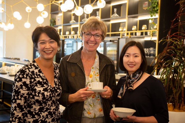
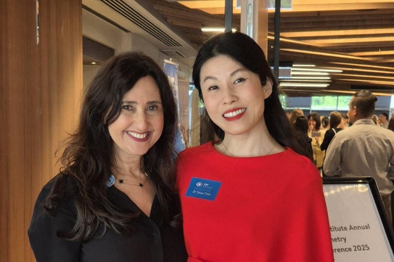
Dr Divya Perumal, Claire McDonald and Inhae Park
Fellow Aucklander Dr Sophie Hill’s presentation acknowledged the increased number of patients receiving often sight-saving intravitreal injections for macular degeneration with associated transient IOP spikes. There is a tricky balance between needle gauge, reportedly a factor in patient experience, and observed reflux, which will blunt the IOP rise. Protocols to aid this balance are currently being developed.
As always, presenters were a mix of ophthalmologists and optometrists. Wellington Hospital optometrist Inhae Park gave a great overview of secondary glaucomas, but will perhaps now be remembered more for her account of being too short to fit into Dr Gale’s inverter! UoA optometrist Dr Hannah Kersten got us thinking about the goal of early detection. Prior to ganglion cell apoptosis might be the best time to treat, before vision loss, but how can we see it happening? Multifocal pattern electroretinogram (ERG) may be useful but has yet to be established as a routine tool. Optometrists Robert Ng, Michelle Jefferies and Alex Petty presented a range of interesting glaucoma cases for discussion, always an engaging session.
The day concluded with research updates from two Gordon Sanderson scholarship recipients, Dr Pratik Chandra and Dr Aqeeda Singh, together with the Frederich recipients, along with Frederich Kurtstein Anthonsen Trust scholar Chris Mayo, whose research project attempted to design a glaucoma risk calculator using artificial intelligence. Dr Chandra offered us some tips to improve treatment adherence, including personalisation and using printed material, phone calls and digital reminders, while participating in the Dunedin Study offered Dr Singh the opportunity to measure glaucoma prevalence in a group of Kiwi 45-year-olds, revealing it to be 0.79%.
A thoroughly worthwhile day, which I recommend adding to your 2023 CPD calendar.
Claire McDonald is an optometrist working in private practice in Warkworth. In 2017 she completed the University of Auckland (UoA) advanced glaucoma care credentialing course, supervised by Dr Hussain Patel. Until recently she worked part time with ADHB glaucoma patients in the UoA clinic.