


Ophthalmologists at New York Eye and Ear Infirmary of Mount Sinai, USA, have demonstrated that sequential optical coherence tomography angiography (OCT-A) can highlight sickle cell disease patients who are at risk of developing retinopathy.
Publishing in Biomedical Optics Express, the study’s authors described using OCT-A to image patients 10 times over 10 minutes, repeating this an hour later. By overlaying the images, researchers could observe retinal capillaries that appeared to ‘flicker’, indicating interruption of blood flow. Of the 27 subjects, the 14 controls’ capillaries remained mostly open, whereas the sickle cell patients who were not receiving hydroxyurea treatment showed significant flickering, indicating the risk of focal vascular stroke and permanent blindness. OCT-A of the retinas of sickle cell patients receiving treatment showed good blood flow, showing that their medication was effective, said researchers.
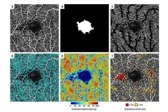
A: Contrast-stretched full vascular slab OCT-A; B: Manual segmentation of foveal avascular zone; C: Full vascular slab OCT-A after removal of non-capillary blood vessels; D: Parafoveal capillary segmentation highlighted in cyan; E: Parafoveal capillary density map with non-capillary blood vessels indicated in white due to the exclusion from density computation; F: Corresponding deviation map. Areas with parafoveal capillary density below 5% and 1% of the normal distribution are indicated in yellow and red, respectively
"Our work can be a game-changer for sickle cell patients, especially for those who have no symptoms of retinopathy. It can lead to earlier diagnosis of retinal issues and prevention of irreversible blindness. Without this technology, it's impossible to judge their eye condition until patients report vision loss, when it's too late," said co-author Professor Richard Rosen, chief of retina services for the Mount Sinai Health System.